Women Day OFFER :GET 50% Discount on Apparel, Shoes, Glares, Sacarfs, Mobile cover etc use the CODE- INTLWOMEN23# ORDERONLINE NOW
$
2500
$ 1500
(40% off)
zabby51
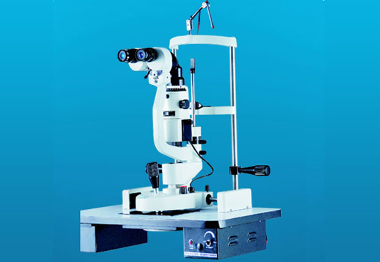
Slit Lamps - 4 Models The slit lamp is essentially a simple and generally under-used piece of equipment. It consists of an illumination system and a binocular observation system, which when correctly aligned will result in a coincidental focus of the slit and microscope. Illumination system Basically a short focus projector projecting an image of the illuminated slit aperture on to the eye. This part of the system should be flexible to allow various sizes and shape of slit beam. Usually a rheostat is incorporated and the lamp house can be rotated. Neutral density, cobalt blue and red free filters are usually available, and occasionally a diffuser and polarizer Using the slit lamp Commence the examination using the 10x eyepieces and the lower powered objective i.e. 1x. Use the lowest voltage setting on the transformer. Select the longest slit length by means of the appropriate lever (17). Adjust the chin rest by means of control 27 so that the patient's eyes are approximately level with the black marker on the side of the head rest. Adjust the height of the slit lamp until the slit beam is centred vertically on the patient's eye. Focus the slit beam on the eye by moving the joystick (1) either towards or away from the patient. Coarse positioning can be effected without using the microscope but critical focussing should be carried out whilst viewing through the microscope. The slit width is varied by rotating either the left hand or right hand knurled control 10. To vary the angle between illumination and microscope use one or other of these same controls as handles. The slit should be set primarily in the vertical position, but any desired inclination can be achieved by means of the ball handle 15 (notches at 45?, 90?, 135?; stops at 0? and 180?). By tripping the latch 11 and tilting the slit lamp column, the beam can be introduced from as much as 20? below the horizontal. This is mainly used for carrying out gonioscopy. For observation by sclerotic scatter or other dissociated forms of examination the centering screw 13 is loosened, so that the slit image can be moved away from the centre of the field of observation. The image is centered again by tightening the screw. Carry out the techniques described below. Slit Lamp - IE-02 Excel Specifications - Binocular Microscope ? Eye Pieces : 10x, 15x ? Objective : 1x, 1.6x ? Total Magnification : 10x, 16x and 24x Illumination Unit ? Slit Image Rotation : 0 to 180? ? Filter Disc : Cobalt Filter, Green Filter, Yellow Filter Natural Density and Open Aperture ? Slit Disphragm Disc : Wedge Shaped Diaphragm Of Infinitely Variable Slit Lengths ? Halogen Lamp : 6 Volt 20 Watts Standard Accessories ? Replacing Lamp ? Plastic Breath Shield ? Testing Rod ? Dust Cover and Fuse ? 15 Eye Pieces
TRy YOU MAY ALSO LIKE THIS COLLECTION
We take this opportunity to introduce ourselves as a leading manufacturer and exporter of goods of highest international standard and to export/supply them at most competitive rates to all parts of the world.


Toll Free
(011 2341 2786) Mon-Sat 10AM - 6PM IST

27, 28 & 93, Lower Ground Floor World Trade Center Near Hotel The Lalit Babar Road, Barakhamba Lane Connaught Place, New Delhi-110001, INDIA


